Cardiac Auscultation Points and Murmurs: Where to Listen and What You Hear
By the DecodeMed Team8 min read
Get the RadioAnato AI app, free:App StoreGoogle PlayAbout RadioAnato AI
There are five classic auscultation points: aortic (2nd right intercostal space at the sternal border), pulmonary (2nd left intercostal space), Erb’s point (3rd left intercostal space), tricuspid (4th–5th left intercostal space at the lower sternal border) and mitral (5th intercostal space in the midclavicular line, the apex). Each murmur is loudest where the blood flow through its valve projects, which is not always over the valve itself.
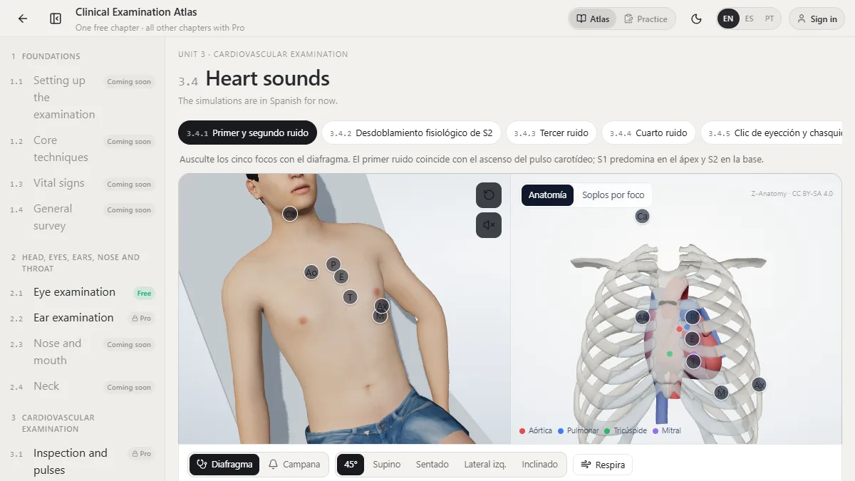
The five auscultation points
| Point | Where | Best heard |
|---|---|---|
| Aortic | 2nd right intercostal space, right sternal border | S2 (A2), aortic stenosis, aortic sclerosis |
| Pulmonary | 2nd left intercostal space, left sternal border | S2 splitting (P2), pulmonary stenosis, flow murmurs |
| Erb’s point | 3rd left intercostal space, left sternal border | Aortic regurgitation, many murmurs at once |
| Tricuspid | 4th–5th left intercostal space, lower left sternal border | Tricuspid regurgitation, ventricular septal defect |
| Mitral (apex) | 5th intercostal space, midclavicular line | S1, S3, S4, mitral regurgitation, mitral stenosis |
Locate the angle of Louis (sternal angle) first: the 2nd rib joins the sternum there, so the 2nd intercostal space is just below it. Count down from there rather than guessing. The apex is felt as the point of maximal impulse; in a dilated heart it moves down and out, and so does the mitral point.
Diaphragm or bell
- Diaphragm (pressed firmly) for high-pitched sounds: S1, S2, aortic and mitral regurgitation, most systolic murmurs.
- Bell (resting lightly) for low-pitched sounds: S3, S4 and the diastolic rumble of mitral stenosis.
The common murmurs
| Murmur | Timing | Loudest at | Radiates to | Clue |
|---|---|---|---|---|
| Aortic stenosis | Mid-systolic, crescendo–decrescendo | Aortic point | Carotids | Slow-rising pulse, soft A2 when severe |
| Mitral regurgitation | Holosystolic (pansystolic) | Apex | Left axilla | Louder with handgrip |
| Aortic regurgitation | Early diastolic, decrescendo | Erb’s point / left sternal border | — | Patient sitting forward, breath held in expiration |
| Mitral stenosis | Mid-diastolic rumble after an opening snap | Apex | — | Bell, patient turned on the left side |
| Tricuspid regurgitation | Holosystolic | Lower left sternal border | Right sternal border | Louder on inspiration |
| Ventricular septal defect | Holosystolic, often harsh | Lower left sternal border | Across the precordium | Small defects can be loud |
| Hypertrophic cardiomyopathy | Mid-systolic | Left sternal border | Rarely to carotids | Louder on standing and with Valsalva |
Manoeuvres that change murmurs
- Inspiration increases right-sided murmurs (tricuspid, pulmonary) because more blood returns to the right heart.
- Handgrip raises afterload: mitral regurgitation, aortic regurgitation and VSD get louder; aortic stenosis and HCM get softer.
- Valsalva and standing reduce preload: almost every murmur gets softer, except hypertrophic cardiomyopathy (louder) and mitral valve prolapse (the click moves earlier).
- Squatting and leg raising increase preload: most murmurs get louder, HCM gets softer.
Grading a murmur
Systolic murmurs are graded 1 to 6 (Levine scale): 1 barely audible, 2 soft but heard at once, 3 moderately loud, 4 loud with a palpable thrill, 5 heard with the stethoscope barely touching, 6 heard with the stethoscope off the chest. Diastolic murmurs are usually graded 1 to 4. Any diastolic murmur, any murmur with a thrill and any murmur with symptoms deserves an echocardiogram.
Practise it on a 3D patient
In the Clinical Examination Atlas you place the stethoscope on each focus of a 3D patient, switch between diaphragm and bell, change the position and try manoeuvres, while the anatomy view on the right shows which valve lies under the point. The practice mode then plays heart sounds and murmurs for you to name.
Study it in RadioAnato AI
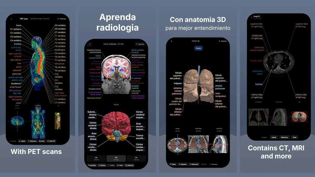
RadioAnato AI, the DecodeMed app, puts 3D anatomy, CT and MRI studies, histology slides and an AI tutor that answers on them in your pocket. Free to download for iPhone, iPad and Android.
See also the best physical exam simulators and, for the electrical side, normal ECG values.
Frequently asked questions
What are the 5 cardiac auscultation points?
Aortic (2nd right intercostal space), pulmonary (2nd left intercostal space), Erb’s point (3rd left intercostal space), tricuspid (lower left sternal border) and mitral (5th intercostal space, midclavicular line).
Where is the aortic stenosis murmur heard best?
At the aortic point, the 2nd right intercostal space, as a crescendo–decrescendo systolic murmur that radiates to the carotids.
How do you tell aortic stenosis from mitral regurgitation?
Aortic stenosis is a mid-systolic ejection murmur at the right upper sternal border radiating to the neck that softens with handgrip; mitral regurgitation is holosystolic at the apex, radiates to the axilla and gets louder with handgrip.
When do you use the bell of the stethoscope?
For low-pitched sounds: S3, S4 and the mid-diastolic rumble of mitral stenosis, best heard at the apex with the patient on the left side.
Get RadioAnato AI, the DecodeMed app
Anatomy, radiology and histology in one app, on the App Store and Google Play.
About RadioAnato AI