The Normal ECG: Waves, Intervals and Normal Values
By the DecodeMed Team7 min read
Get the RadioAnato AI app, free:App StoreGoogle PlayAbout RadioAnato AI
A normal adult ECG shows sinus rhythm at 60–100 bpm, a PR interval of 120–200 ms, a QRS under 120 ms, a QTc under about 450 ms in men and 460 ms in women, an axis between −30° and +90°, an isoelectric ST segment and upright T waves in I, II and V3–V6. Values vary slightly between references.
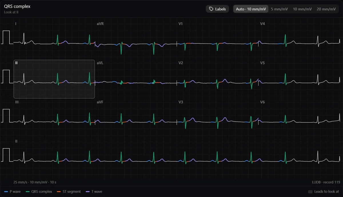
Normal values at a glance
| Measure | Typical normal value (adult) | Where to look |
|---|---|---|
| Heart rate | 60–100 bpm | Rhythm strip |
| P wave | Under 120 ms and under 2.5 mm tall | Lead II, V1 |
| PR interval | 120–200 ms | Lead II |
| QRS duration | Under 120 ms (usually 80–110) | Widest lead |
| QTc | Under about 450 ms (men), 460 ms (women) | Lead II or V5 |
| Axis | −30° to +90° | Limb leads |
| R-wave transition | V3–V4 | Chest leads |
P wave and PR interval
- Sinus P waves are upright in I, II and aVF and inverted in aVR. In V1 the P wave is often biphasic.
- A wide or notched P suggests left atrial enlargement; a tall P in II suggests right atrial enlargement.
- PR over 200 ms is first-degree AV block; under 120 ms raises pre-excitation or a low atrial rhythm.
QRS complex
- Width: 120 ms or more is a bundle branch block, ventricular rhythm or pacing until proven otherwise.
- Small septal q waves are normal in I, aVL, V5 and V6. A Q wave of 40 ms or more, or deeper than a quarter of the R wave, is pathological.
- R-wave progression: the R wave grows from V1 to V6 and equals the S wave around V3–V4.
ST segment, T wave and QT
- ST segment: at the baseline, measured at the J point. A small ST elevation in V2–V3 can be normal, especially in young men.
- T waves: upright in I, II and V3–V6, inverted in aVR; they can be inverted in III, aVL, aVF and V1 in healthy people.
- QT: varies with rate, so correct it (QTc). A QTc over 500 ms carries a high risk of arrhythmia.
- U wave: a small wave after the T, best seen in V2–V3, can be normal.
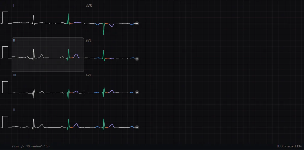
Normal variants that are not disease
- Sinus bradycardia in athletes and during sleep.
- Sinus arrhythmia: the rate rises with inspiration and falls with expiration.
- Early repolarisation: J-point elevation with a notch or slur, common in young people.
- A right axis in children and some slim young adults.
Study it in RadioAnato AI
RadioAnato AI, the DecodeMed app, puts 3D anatomy, CT and MRI studies, histology slides and an AI tutor that answers on them in your pocket. Free to download for iPhone, iPad and Android.
See it on a real tracing
Every value above appears on real tracings in the ECG atlas, with each wave coloured where cardiologists marked it and the intervals measured. To read in order, follow how to learn ECG from scratch; for rate and axis, see heart rate and axis.
Frequently asked questions
What is the normal PR interval?
120 to 200 ms (3 to 5 small squares at 25 mm/s).
What is a normal QRS duration?
Under 120 ms, usually 80 to 110 ms. 120 ms or more is a wide QRS.
What is a normal QTc?
Under about 450 ms in men and 460 ms in women; over 500 ms is high risk. Cut-offs vary slightly between guidelines.
Get RadioAnato AI, the DecodeMed app
Anatomy, radiology and histology in one app, on the App Store and Google Play.
About RadioAnato AI