How to Learn the Brachial Plexus Fast (Roots, Trunks, Cords, Branches)
By the DecodeMed Team8 min read
Get the RadioAnato AI app, free:App StoreGoogle PlayAbout RadioAnato AI
The brachial plexus is built from five roots (C5–T1) that join into three trunks (upper, middle, lower), split into anterior and posterior divisions, regroup into three cords named by their position around the axillary artery (lateral, posterior, medial) and end as five terminal nerves: musculocutaneous, axillary, radial, median and ulnar. Learn it in that order, “Randy Travis Drinks Cold Beer”, and then draw it until you can do it in one minute.
The five parts, in order
| Part | What forms | Where it lies |
|---|---|---|
| Roots | Ventral rami of C5, C6, C7, C8, T1 | Between anterior and middle scalene |
| Trunks | Upper (C5–C6), middle (C7), lower (C8–T1) | Posterior triangle of the neck |
| Divisions | Each trunk splits into anterior and posterior | Behind the clavicle |
| Cords | Lateral (anterior divisions of upper and middle), medial (anterior of lower), posterior (all posterior divisions) | Axilla, around the 2nd part of the axillary artery |
| Branches | Musculocutaneous, axillary, radial, median, ulnar | Into the arm |
A rule that removes half the memorising: anterior divisions supply flexors, posterior divisions supply extensors. That is why the posterior cord gives the axillary and radial nerves (extensors of the shoulder, elbow and wrist), and the lateral and medial cords give the musculocutaneous, median and ulnar nerves (flexors).
The M shape
In front of the axillary artery, the musculocutaneous, median and ulnar nerves form a letter M: the lateral cord gives the musculocutaneous nerve and the lateral root of the median nerve, the medial cord gives the medial root of the median nerve and the ulnar nerve. Find this M first in a dissection or a 3D model; everything else hangs off it.
Branches you must know
| From | Nerve | Supplies |
|---|---|---|
| Roots | Dorsal scapular (C5) | Rhomboids, levator scapulae |
| Roots | Long thoracic (C5–C7) | Serratus anterior |
| Upper trunk | Suprascapular | Supraspinatus, infraspinatus |
| Lateral cord | Lateral pectoral | Pectoralis major |
| Medial cord | Medial pectoral; medial cutaneous nerves of arm and forearm | Pectoralis minor and major; skin |
| Posterior cord | Upper and lower subscapular, thoracodorsal (STAR: subscapulars, thoracodorsal, axillary, radial) | Subscapularis, teres major, latissimus dorsi |
Classic injuries
| Injury | Typical cause | What you see |
|---|---|---|
| Erb–Duchenne (C5–C6) | Head pushed away from the shoulder, difficult birth | “Waiter’s tip”: arm adducted, internally rotated, elbow extended |
| Klumpke (C8–T1) | Arm pulled upward | Claw hand, small hand muscles weak; Horner syndrome if T1 is torn near the cord |
| Long thoracic nerve | Mastectomy, carrying loads on the shoulder | Winged scapula |
| Axillary nerve | Fracture of the surgical neck, shoulder dislocation | Weak abduction, numb “regimental badge” area |
| Radial nerve | Mid-shaft humerus fracture | Wrist drop |
| Median nerve | Carpal tunnel syndrome, supracondylar fracture | Thenar weakness, numb lateral three and a half fingers |
| Ulnar nerve | Medial epicondyle injury | Claw of the ring and little fingers, weak finger abduction |
A 20-minute method
- Draw it from roots to branches, out loud, three times.
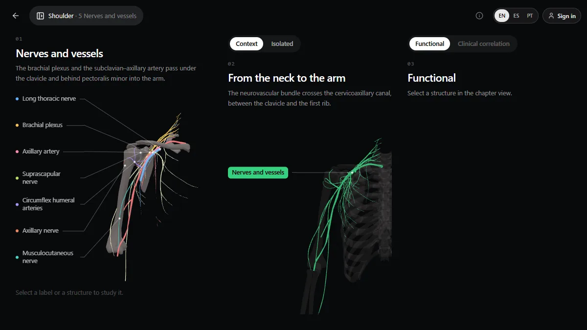
- See it in 3D. In the shoulder lesson of the 3D anatomy atlas, the “Nerves and vessels” chapter shows the plexus with the axillary artery, the long thoracic, suprascapular, axillary and musculocutaneous nerves labelled, and its path from the neck to the arm. Turn it until you can name each label before clicking it.
- Test yourself on injuries: for each nerve, name the cause, the muscle that fails and the sign.
- Ask the AI tutor in RadioAnato AI to quiz you on a random branch, and make flashcards of what you miss.
Study it in RadioAnato AI
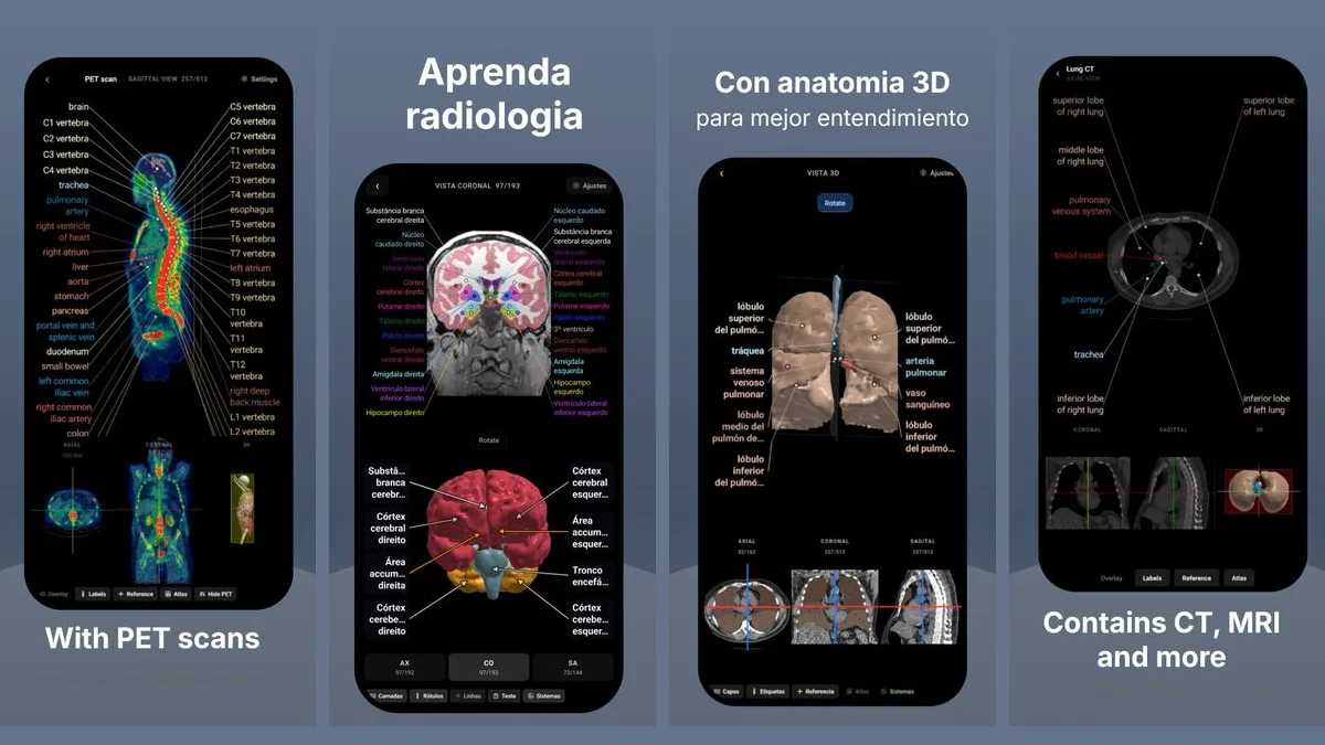
RadioAnato AI, the DecodeMed app, puts 3D anatomy, CT and MRI studies, histology slides and an AI tutor that answers on them in your pocket. Free to download for iPhone, iPad and Android.
For a full study routine see how to study anatomy in medical school and the best apps to learn anatomy.
Frequently asked questions
What are the 5 terminal branches of the brachial plexus?
Musculocutaneous, axillary, radial, median and ulnar nerves.
What is the mnemonic for the brachial plexus?
“Randy Travis Drinks Cold Beer”: Roots, Trunks, Divisions, Cords, Branches. For the posterior cord branches, STAR: upper and lower Subscapular, Thoracodorsal, Axillary, Radial.
Why are the cords called lateral, medial and posterior?
Because of their position around the second part of the axillary artery, behind pectoralis minor.
What does an Erb palsy look like?
A C5–C6 injury: the arm hangs adducted and internally rotated with the elbow extended and the forearm pronated, the “waiter’s tip” position.
Get RadioAnato AI, the DecodeMed app
Anatomy, radiology and histology in one app, on the App Store and Google Play.
About RadioAnato AI