Coronary Artery Anatomy: Branches, Territories and Dominance
By the DecodeMed Team7 min read
Get the RadioAnato AI app, free:App StoreGoogle PlayAbout RadioAnato AI
Two coronary arteries arise from the aortic sinuses just above the aortic valve. The left main coronary artery splits into the left anterior descending (LAD), which supplies the anterior wall, the apex and the anterior two-thirds of the septum, and the circumflex, which supplies the lateral wall. The right coronary artery (RCA) supplies the right ventricle and, in most people, the inferior wall through the posterior descending artery.
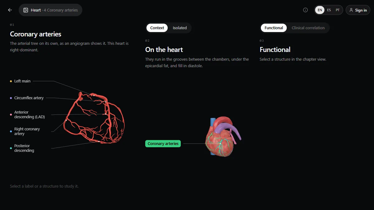
The arteries and their branches
| Artery | Course | Main branches | Supplies |
|---|---|---|---|
| Left main | From the left aortic sinus, behind the pulmonary trunk, 1–2 cm long | LAD and circumflex (sometimes a ramus intermedius) | Most of the left ventricle |
| Left anterior descending (LAD) | Anterior interventricular groove to the apex | Diagonals, septal perforators | Anterior wall, apex, anterior two-thirds of the septum |
| Circumflex | Left atrioventricular groove, around to the back | Obtuse marginals | Lateral wall, part of the posterior wall |
| Right coronary (RCA) | Right atrioventricular groove, around to the back | Conus, SA nodal (often), acute marginal, AV nodal, posterior descending | Right ventricle, inferior wall, posterior third of the septum, SA and AV nodes |
Coronary dominance
Dominance is defined by which artery gives the posterior descending artery (PDA). About 70–80% of people are right-dominant (PDA from the RCA), around 10% left-dominant (from the circumflex) and the rest co-dominant. It matters clinically: in a left-dominant heart, a circumflex occlusion can cause an inferior infarct, and a left main occlusion threatens almost the whole left ventricle.
The nodes
- Sinoatrial (SA) node artery: from the RCA in about 60% of people, from the circumflex in the rest.
- Atrioventricular (AV) node artery: from the dominant artery, so usually the RCA. That is why inferior infarcts often cause bradycardia and AV block.
From artery to ECG leads
| Wall | ECG leads | Usual culprit artery |
|---|---|---|
| Anterior / anteroseptal | V1–V4 | LAD |
| Lateral | I, aVL, V5–V6 | Circumflex or a diagonal of the LAD |
| Inferior | II, III, aVF | RCA (or circumflex if left-dominant) |
| Posterior | ST depression in V1–V3, tall R; ST elevation in V7–V9 | Circumflex or RCA |
| Right ventricle | ST elevation in V4R | Proximal RCA |
Cardiac veins in one line
The veins run with the arteries in the same grooves: the great cardiac vein with the LAD and then the circumflex, the middle cardiac vein with the PDA, the small cardiac vein with the RCA, and all drain into the coronary sinus, which opens into the right atrium.
See it in 3D and on CT
The heart lesson of the 3D anatomy atlas has a “Coronary arteries” chapter that shows the tree on its own, as an angiogram shows it, with the left main, circumflex, LAD, RCA and PDA labelled, and the same arteries on the heart beside it. The next chapter does the same for the cardiac veins. The radiology atlas has a heart CT to see the heart and great vessels on real slices.
Study it in RadioAnato AI
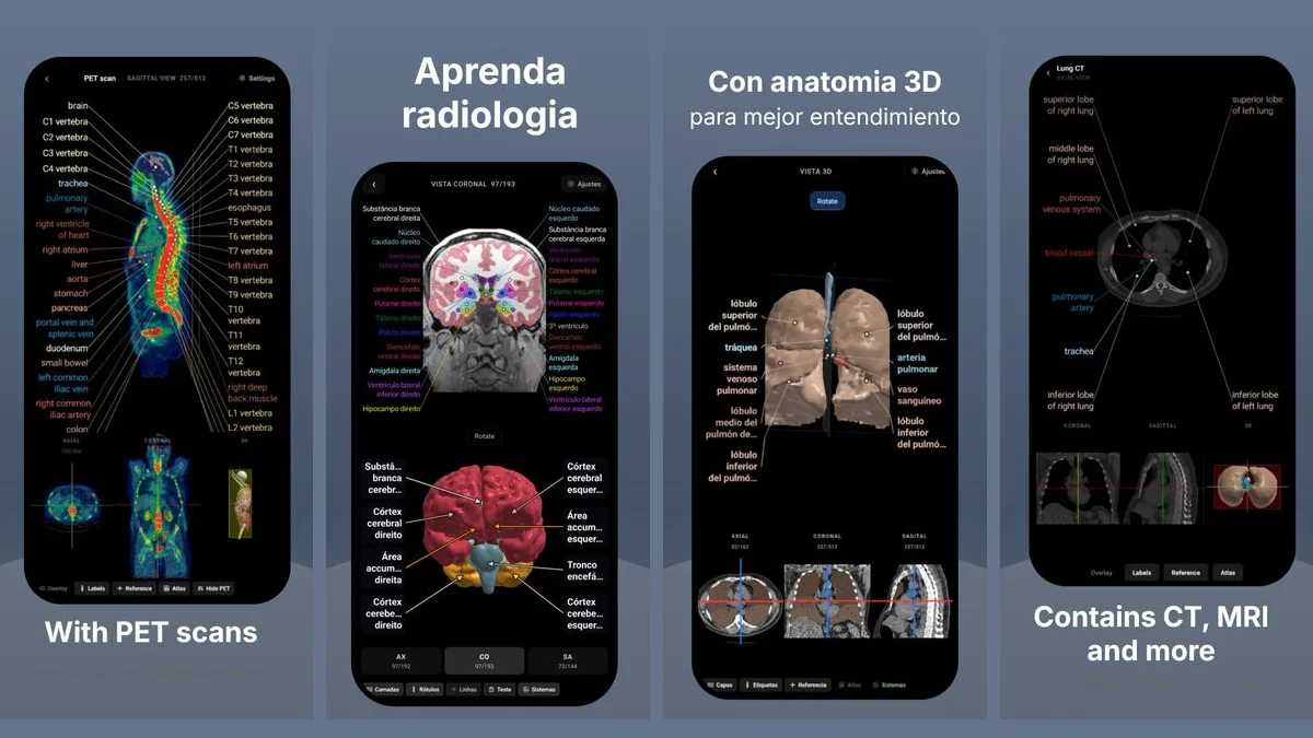
RadioAnato AI, the DecodeMed app, puts 3D anatomy, CT and MRI studies, histology slides and an AI tutor that answers on them in your pocket. Free to download for iPhone, iPad and Android.
Connect it to the ECG with how to learn ECG from scratch and to imaging with chest CT anatomy.
Frequently asked questions
What are the main coronary arteries?
The left main coronary artery, which divides into the left anterior descending and the circumflex, and the right coronary artery.
What does the LAD supply?
The anterior wall of the left ventricle, the apex and the anterior two-thirds of the interventricular septum. Its occlusion causes an anterior infarct seen in V1–V4.
What is right coronary dominance?
When the posterior descending artery comes from the right coronary artery, which is the case in about 70–80% of people.
Which artery supplies the AV node?
The dominant artery, usually the right coronary artery, which is why inferior infarcts often cause AV block.
Get RadioAnato AI, the DecodeMed app
Anatomy, radiology and histology in one app, on the App Store and Google Play.
About RadioAnato AI