Chest CT Anatomy: The Mediastinum and Lungs, Level by Level
Chest CT anatomy is easiest to learn as six axial levels, read from top to bottom in two windows. The mediastinal window shows the heart, the great vessels and the lymph nodes; the lung window shows the airways, the lobes and the fissures. At each level, name the same few structures.
Two windows, two questions
| Window | Typical setting | What you study |
|---|---|---|
| Mediastinal (soft tissue) | W 350 to 400, L 40 to 50 | Heart, great vessels, oesophagus, lymph nodes, chest wall |
| Lung | W 1500, L −600 | Trachea and bronchi, lung parenchyma, fissures, pleura |
| Bone | W 2000, L 500 | Ribs, sternum, spine |
Why these numbers work is explained in Hounsfield units and CT windows.
Six levels of the mediastinum
- 1. Above the aortic arch: the trachea in the midline with the oesophagus behind it, the two brachiocephalic veins in front, and the three arch branches: brachiocephalic trunk, left common carotid and left subclavian artery.
- 2. Aortic arch: the arch crosses from front right to back left, to the left of the trachea. The superior vena cava lies on the right.
- 3. Aortopulmonary window: the space under the arch and above the left pulmonary artery. It holds fat and lymph nodes. The azygos vein arches forward to join the superior vena cava at about this level.
- 4. Carina and pulmonary arteries: the trachea divides into the main bronchi. The pulmonary trunk divides: the right pulmonary artery passes behind the ascending aorta and the superior vena cava; the left one arches over the left main bronchus.
- 5. Left atrium: the most posterior chamber, directly in front of the oesophagus, receiving the pulmonary veins. The aortic root lies in the centre.
- 6. Four chambers: the right ventricle is the most anterior chamber, behind the sternum. The left ventricle forms the left border and the right atrium the right border.
The lungs in the lung window
- Lobes: three on the right (upper, middle, lower) and two on the left (upper, which includes the lingula, and lower).
- Fissures: the oblique fissure on both sides separates the lower lobe from the rest; the horizontal fissure exists only on the right, between the upper and middle lobes. On CT a fissure is a thin line or a band with no vessels.
- Bronchi and arteries travel together to the centre of each segment; the veins run separately, between segments.
- The pleura is not seen as a separate layer when it is normal.
Normal sizes worth remembering
- Ascending aorta: under 4 cm.
- Pulmonary trunk: up to about 3 cm, and no wider than the ascending aorta beside it.
- Mediastinal lymph nodes: up to 1 cm in short axis.
Learn it on a labelled chest CT
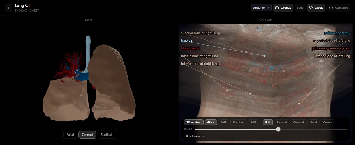
The lung and heart CT studies in the free radiology atlas show the lobes, the trachea and the pulmonary arteries and veins as labelled 3D models next to the slices. Scroll from the arch to the diaphragm naming the six levels, first with the labels and then without. The abdomen follows the same logic: see how to read an abdominal CT.
A search pattern for the whole scan
- Mediastinal window: thyroid, great vessels, heart and pericardium, lymph nodes, oesophagus, chest wall and axillae.
- Lung window: trachea and bronchi, each lobe in turn, fissures and pleura.
- Bone window: ribs, sternum, spine.
- Do not forget the edges: the lower neck and the upper abdomen are on the scan too.
Frequently asked questions
What are the mediastinal and lung windows on chest CT?
They are two ways of displaying the same slices. The mediastinal window (width about 350 to 400 HU, level 40 to 50 HU) shows soft tissues such as the heart, vessels and lymph nodes. The lung window (width 1500 HU, level −600 HU) shows the airways and lung parenchyma.
Which heart chamber is most anterior on CT?
The right ventricle, which lies directly behind the sternum. The left atrium is the most posterior chamber, in front of the oesophagus.
What is the aortopulmonary window?
The small space between the underside of the aortic arch and the top of the left pulmonary artery. It normally contains fat, small lymph nodes and the left recurrent laryngeal nerve.
Get the DecodeMed app
Anatomy, radiology and histology in one app, on the App Store and Google Play.