Hounsfield Units and CT Windows Explained, with a Value Table
A CT image is a map of density. Each pixel has a number in Hounsfield units (HU): water is 0, air is −1000 and dense bone is about +1000. Windowing chooses which part of that scale is spread across the greys on the screen, which is why the same slice looks different in a lung window and a bone window.
The Hounsfield scale
| Tissue | Approximate HU | How it looks |
|---|---|---|
| Air | −1000 | Black |
| Lung | −700 to −900 | Almost black |
| Fat | −50 to −120 | Dark grey |
| Water, simple fluid | 0 to 20 | Grey |
| White matter | 20 to 30 | Grey |
| Grey matter | 35 to 45 | Slightly lighter grey |
| Muscle and solid organs | 40 to 70 | Light grey |
| Acute blood | 50 to 90 | Bright |
| Calcification | Over 130 | White |
| Cortical bone | About 1000 and above | White |
The numbers are approximate and change with the scanner and the energy used. Intravenous contrast raises the HU of whatever it reaches, which is how enhancement is measured: a rise of more than about 20 HU between the unenhanced and enhanced scan means the tissue enhances.
Window width and window level
The scanner records thousands of HU values, but a screen shows a limited number of greys. The window level (L) is the HU value placed at mid-grey; choose it close to the tissue you want to study. The window width (W) is the range of HU spread from black to white. A narrow window gives high contrast between similar tissues; a wide one shows very different tissues together.
The displayed range is L − W/2 to L + W/2. With W 400 and L 40, everything below −160 HU is black and everything above +240 HU is white.
The windows you will use
| Window | Width (W) | Level (L) | Used for |
|---|---|---|---|
| Brain | 80 | 40 | Grey and white matter, haemorrhage |
| Stroke | 40 | 40 | Early loss of grey-white differentiation |
| Subdural | 200 | 80 | Thin blood next to the skull |
| Soft tissue (abdomen, mediastinum) | 350 to 400 | 40 to 50 | Organs, vessels, nodes |
| Lung | 1500 | −600 | Lung parenchyma, airways, free air |
| Bone | 2000 | 500 | Cortex, trabeculae, fractures |
Each department uses slightly different presets, so treat these as typical values.
How to use HU when you read a scan
- Measure, do not guess. Place a region of interest on a lesion: near 0 HU is fluid, negative values mean fat, 50 to 90 HU in the unenhanced scan suggests fresh blood.
- Change the window on purpose. Look for free air in the lung window and for fractures in the bone window.
- Beware of small structures. A thin lesion averages its density with its neighbours (partial volume), so its HU may mislead.
- Compare before and after contrast in the same place to decide whether something enhances.
See it on a real scan
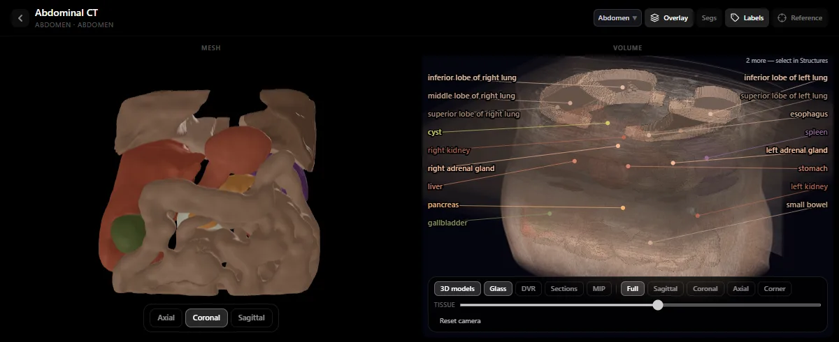
The free radiology atlas lets you scroll real CT studies with the organs labelled, which is the fastest way to connect these numbers to what you see. Then follow a full method in how to read an abdominal CT.
Frequently asked questions
What are Hounsfield units?
They are the scale CT uses to express how much a tissue attenuates X-rays. Water is defined as 0 HU and air as −1000 HU; fat is negative, soft tissues are between about 20 and 70 HU, and bone is several hundred to more than 1000 HU.
What is the difference between window width and window level?
The level is the HU value shown as mid-grey, so it sets the brightness and is chosen near the tissue of interest. The width is the range of HU shown from black to white, so it sets the contrast.
What window is used for the lungs on CT?
A wide window centred on low density, typically a width of 1500 HU and a level of −600 HU.
Get the DecodeMed app
Anatomy, radiology and histology in one app, on the App Store and Google Play.