Liver Segments on CT: How to Identify the Couinaud Segments
The Couinaud classification divides the liver into eight segments, each with its own portal inflow and venous outflow. On CT you find them with two sets of landmarks: the three hepatic veins divide the liver from side to side, and the plane of the right and left portal veins divides it into upper and lower segments.
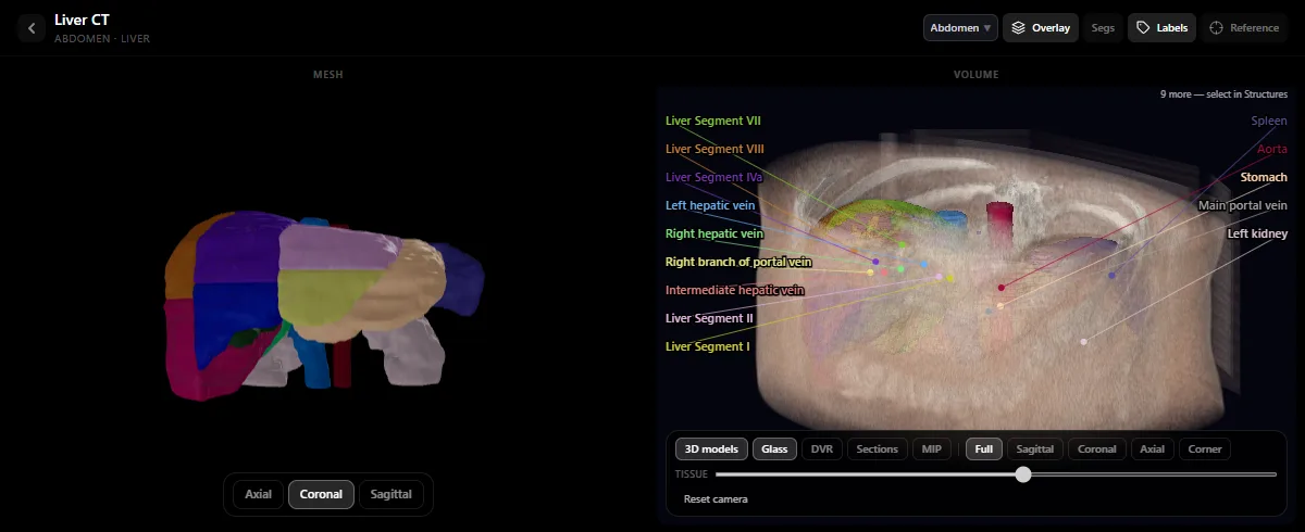
Why eight segments
Each segment receives its own branch of the portal vein, hepatic artery and bile duct, and drains towards a hepatic vein. A surgeon can therefore remove one segment without damaging its neighbours. That is why reports state the segment in which a lesion lies.
The two sets of landmarks
- Middle hepatic vein: separates the right liver from the left liver. Its plane runs from the gallbladder fossa to the inferior vena cava.
- Right hepatic vein: divides the right liver into an anterior part (segments V and VIII) and a posterior part (segments VI and VII).
- Left hepatic vein and falciform ligament: separate the medial segment IV from the lateral segments II and III.
- Portal vein plane: the right and left portal branches separate the upper segments (II, IVa, VII, VIII) from the lower ones (III, IVb, V, VI).
The eight segments
| Segment | Position |
|---|---|
| I | Caudate lobe: posterior, between the inferior vena cava and the portal vein |
| II | Left lateral, above the portal plane |
| III | Left lateral, below the portal plane |
| IV | Left medial: IVa above and IVb below the portal plane |
| V | Right anterior, below the portal plane |
| VI | Right posterior, below the portal plane |
| VII | Right posterior, above the portal plane |
| VIII | Right anterior, above the portal plane |
Seen from the front, the segments are numbered clockwise, starting with the caudate lobe at the centre.
Reading the segments on axial slices
- Above the portal bifurcation, where the three hepatic veins reach the vena cava: from the patient’s left to right you cross II, IVa, VIII and VII, with a hepatic vein between each pair.
- At the portal bifurcation: the left portal vein marks the change from II to III on the left; the right portal vein marks the change from the upper to the lower right segments. The caudate lobe lies just behind, in front of the vena cava.
- Below the portal bifurcation: from left to right, III, IVb, V and VI. The gallbladder lies on the boundary between IVb and V.
The portal venous phase is the easiest one to use, because the hepatic and portal veins are bright. Follow each vein through several slices; do not judge from one image.
See the segments in 3D
The Liver CT study in the free radiology atlas shows the segments as colour-coded 3D models, together with the three hepatic veins and the portal vein, so you can turn the liver and see why the veins are the boundaries. Then return to the slices and find the same veins. The method for the rest of the abdomen is in how to read an abdominal CT.
Keep in mind
- The boundaries are planes, not visible lines. They are extrapolated from the veins.
- Variants are common, especially in the hepatic veins and the right portal branches, so real livers do not always follow the diagram.
- Segment I drains directly into the vena cava and receives blood from both portal branches, which is why it behaves differently in some diseases.
Frequently asked questions
How many segments does the liver have?
Eight in the Couinaud classification, numbered I to VIII. Segment IV is often divided into IVa (upper) and IVb (lower).
How do you identify liver segments on CT?
Use the three hepatic veins to divide the liver from side to side and the plane of the right and left portal veins to divide upper from lower segments. Above the portal plane the order from the patient’s left to right is II, IVa, VIII, VII; below it, III, IVb, V, VI.
Which vein separates the right and left liver?
The middle hepatic vein. Its plane runs from the gallbladder fossa to the inferior vena cava.
Get the DecodeMed app
Anatomy, radiology and histology in one app, on the App Store and Google Play.