How to Read an Abdominal CT: A Step-by-Step Method
Read an abdominal CT the same way every time: check what kind of study it is, look at it in three windows, then go through the organs in a fixed order from the lung bases to the pelvis. A fixed order is what stops you from missing the finding you were not looking for.
Before you look at the organs
- Who and why: the patient’s name, the date and the clinical question.
- Contrast and phase: none, arterial (about 30 seconds after injection, bright aorta), portal venous (about 70 seconds, bright portal and hepatic veins; the standard phase) or delayed (minutes later, contrast in the ureters and bladder). Check for oral contrast too.
- Orientation: axial slices are shown as if you stood at the patient’s feet, so the patient’s right side is on the left of the screen.
Use three windows
| Window | Typical setting | What to look for |
|---|---|---|
| Soft tissue | W 400, L 40 | Organs, vessels, bowel wall, fluid |
| Lung | W 1500, L −600 | Lung bases and free air in the abdomen |
| Bone | W 2000, L 500 | Spine, ribs, pelvis |
Settings vary between departments. The reasoning behind them is in Hounsfield units and CT windows.
A fixed order, from top to bottom
- 1. Lung bases and pleura (lung window): consolidation, effusion, nodules.
- 2. Liver: size, contour, density and focal lesions; then the hepatic and portal veins.
- 3. Gallbladder and bile ducts: stones, wall thickening. The common bile duct normally measures up to about 6 mm.
- 4. Spleen: size and focal lesions.
- 5. Pancreas: follow it from head to tail; look at the duct and the surrounding fat.
- 6. Adrenal glands: thin limbs shaped like a Y or a lambda; any nodule.
- 7. Kidneys, ureters and bladder: stones, hydronephrosis, masses, symmetrical enhancement.
- 8. Stomach and bowel: follow them from the stomach to the rectum and find the appendix. Usual upper limits are 3 cm for small bowel, 6 cm for colon and 9 cm for the caecum.
- 9. Vessels: aorta (under 3 cm), inferior vena cava, portal and mesenteric vessels.
- 10. Lymph nodes, peritoneum and retroperitoneum: enlarged nodes, free fluid, fat stranding, collections.
- 11. Pelvis: bladder, uterus and ovaries or prostate, rectum.
- 12. Bones and abdominal wall (bone window): fractures, focal lesions, hernias.
Landmarks that tell you where you are
- Coeliac trunk: about T12.
- Superior mesenteric artery: about L1.
- Renal hila: L1 to L2.
- Aortic bifurcation: L4, at the level of the iliac crests.
Practise on a labelled scan
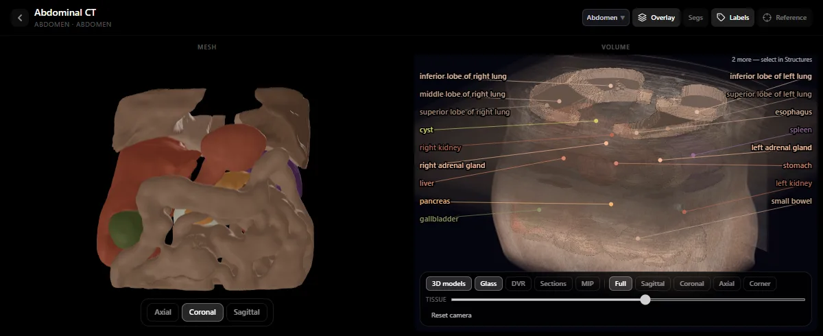
Open the abdominal CT in the free radiology atlas and scroll one organ at a time with the labels on, then hide them and name what you see. Seeing the same organs as 3D models makes the cross-sections easier to hold in your head. For the liver, continue with liver segments on CT.
Common mistakes
- Stopping at the first finding. Finish the whole list every time.
- Staying in the soft-tissue window. Free air and bone lesions hide there.
- Reading without the clinical question, or without comparing with earlier studies.
This is a method for learning. It does not replace the radiologist’s report.
Frequently asked questions
How do you read an abdominal CT systematically?
Check the patient, the question and the contrast phase; review the study in soft-tissue, lung and bone windows; then go through the organs in the same order every time, from the lung bases down to the pelvis, and finish with the bones.
Which CT phase is used for the abdomen?
Most abdominal CTs are read in the portal venous phase, about 70 seconds after injection. A non-contrast scan is used for stones, an arterial phase is added for bleeding and vascular questions, and a delayed phase for the urinary tract.
Which side is the patient’s right on a CT?
On axial slices the patient’s right is on the left of the screen, because the image is viewed from the feet. The liver should therefore appear on the left of the image.
Get the DecodeMed app
Anatomy, radiology and histology in one app, on the App Store and Google Play.