T1 vs T2 MRI: How to Tell Them Apart in Seconds
To tell T1 from T2, look at fluid first. Cerebrospinal fluid, urine or joint fluid is dark on T1 and bright on T2. Then check fat, which is bright on T1, and in the brain check the grey and white matter: on T1 white matter is the lighter of the two, on T2 it is the darker.
The three-step check
- 1. Fluid. Dark fluid means T1 (or FLAIR, see below). Bright fluid means T2.
- 2. Fat. Subcutaneous fat and bone marrow are bright on T1. They are also fairly bright on most T2 images, so fat alone does not separate the two.
- 3. Brain. On T1 the image looks like the anatomy: white matter light, grey matter darker. On T2 the contrast is reversed.
What is bright and what is dark
| Tissue | T1 | T2 | FLAIR |
|---|---|---|---|
| Fluid (CSF, urine, cysts) | Dark | Bright | Dark |
| Fat | Bright | Intermediate to bright | Intermediate to bright |
| White matter | Lighter than grey matter | Darker than grey matter | Darker than grey matter |
| Grey matter | Darker than white matter | Lighter than white matter | Lighter than white matter |
| Oedema, most lesions | Dark | Bright | Bright |
| Air, cortical bone | Dark | Dark | Dark |
The other sequences you will meet
- FLAIR: a T2-type image with the cerebrospinal fluid suppressed. Lesions beside the ventricles and in the cortex stand out because the bright fluid is gone.
- T1 with gadolinium: a T1 image in which vessels and tissues that take up contrast become bright. Compare it with the T1 taken before contrast.
- Fat-suppressed images (STIR, fat-sat): the fat turns dark, so fluid and oedema stand out. They are the workhorse of musculoskeletal MRI.
- DWI and ADC: restricted diffusion is bright on DWI and dark on the ADC map, the pattern of an acute infarct.
- Gradient echo and SWI: blood products and calcium appear black and look larger than they are.
Why some things are bright on T1
Most disease is dark on T1 and bright on T2, because disease usually means more water. A lesion that is bright on T1 before contrast is therefore informative. The usual causes are fat, subacute blood (methaemoglobin), fluid rich in protein and melanin.
TR and TE, in one line each
- T1-weighted: short repetition time (TR) and short echo time (TE).
- T2-weighted: long TR and long TE.
- Proton density: long TR and short TE.
Practise on real studies
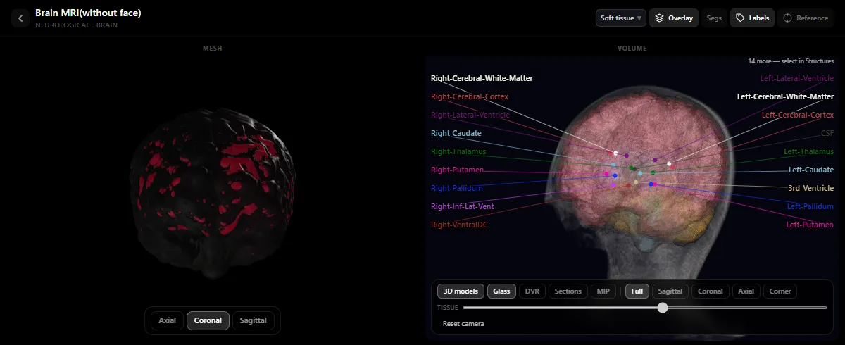
Open a brain MRI in the free radiology atlas, find the lateral ventricles and decide which sequence you are looking at before you read anything else. The labelled structures let you check yourself: caudate, putamen, thalamus and ventricles are the landmarks to learn first. For CT, see Hounsfield units and CT windows.
Frequently asked questions
How do you tell T1 from T2 on MRI?
Look at fluid. Cerebrospinal fluid is dark on T1 and bright on T2. In the brain, white matter is lighter than grey matter on T1 and darker than grey matter on T2.
Is FLAIR a T1 or a T2 sequence?
FLAIR is a T2-type sequence in which the signal of free fluid is suppressed, so cerebrospinal fluid is dark while oedema and most lesions stay bright.
What is bright on T1?
Fat, subacute blood, protein-rich fluid, melanin and anything that takes up gadolinium.
Get the DecodeMed app
Anatomy, radiology and histology in one app, on the App Store and Google Play.