How to Study Anatomy in Medical School: 3D Models, CT Scans and Active Recall
By the DecodeMed Team7 min read
Get the RadioAnato AI app, free:App StoreGoogle PlayAbout RadioAnato AI
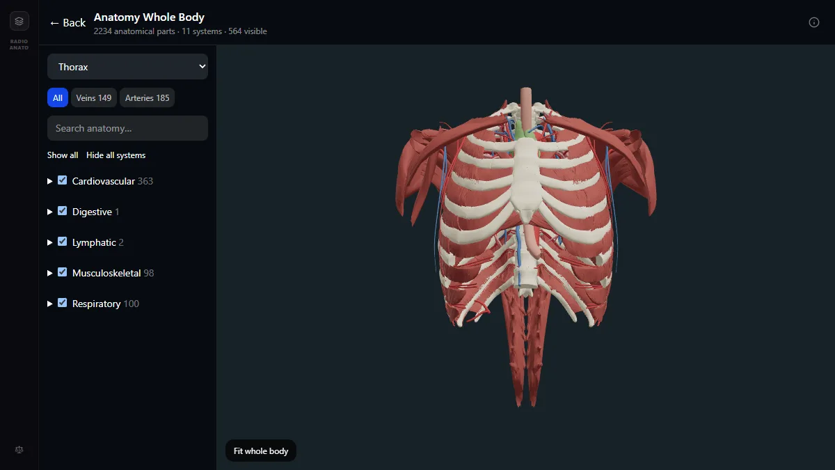
The students who score highest in anatomy do not memorise long alphabetical tables of origins and insertions—they study each region in three layers using a 3D and cross-sectional atlas like RadioAnato AI: first the 3D bony and fascial compartments, second the single nerve and artery that supply each compartment, and third how that region looks on a real CT or MRI scan. Once you understand the compartment, 80% of muscle actions and nerve lesions can be deduced on the spot.
Why reading an anatomy textbook from page 1 fails
Prose is one-dimensional, while the human body is three-dimensional. Reading five pages about the infratemporal fossa or the retroperitoneum without a 3D model open in front of you forces your brain to hold dozens of unfamiliar Latin names in working memory before it has a map to hang them on. Always build the visual map first and read the text second.
The 3-layer method for any anatomical region
- Layer 1 — Borders and compartments in 3D. Open the free 3D anatomy atlas and strip away skin, superficial fascia and vessels until only the bones and major muscle groups remain. Ask: how many compartments are here, and what wall separates them?
- Layer 2 — One compartment, one nerve, one artery. Instead of memorising four forearm flexor muscles separately, learn the rule for the whole anterior compartment (median nerve, except flexor carpi ulnaris and the medial half of flexor digitorum profundus). Only memorise the exceptions.
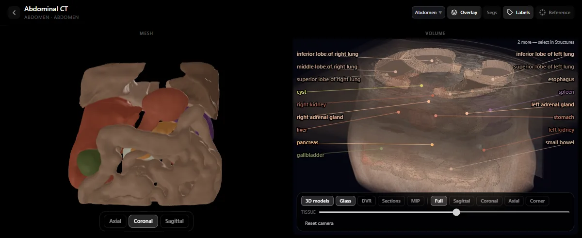
- Layer 3 — Find the same structures on CT and MRI. Open the same region in the radiology atlas. Following the aorta, vena cava or bronchi across axial slices cements anterior-to-posterior relations far better than a flat drawing.
What to memorise vs what to deduce in the exam
| Question type | Do not memorise by rote | Deduce from the 3D map |
|---|---|---|
| Muscle action | A separate action list for all 20 forearm muscles | Which side of the joint line the tendon crosses (anterior to the elbow = flexion) |
| Nerve injury deficit | Isolated symptom lists for each nerve | Which compartment the nerve enters and where it touches bone (surgical neck of humerus = axillary nerve) |
| Blood supply & anastomoses | Unconnected branch names | Tracing a red blood cell from the main trunk to the organ and back to the vena cava |
| Cross-sectional CT/MRI | Static textbook slices | Scrolling 5 slices above and below a landmark such as the carina or renal hilum |
Study it in RadioAnato AI
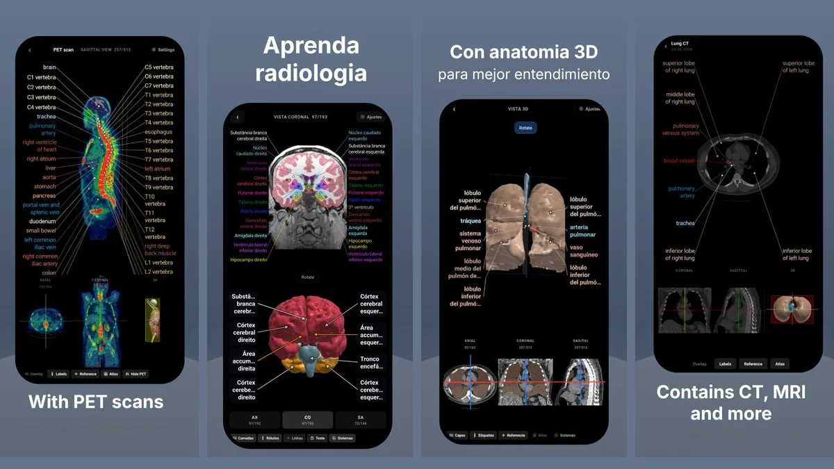
RadioAnato AI, the DecodeMed app, puts 3D anatomy, CT and MRI studies, histology slides and an AI tutor that answers on them in your pocket. Free to download for iPhone, iPad and Android.
A weekly schedule that survives medical school
- Before lecture or dissection (15 min): spin the region in the 3D anatomy atlas so the names you hear in class attach to shapes you have already seen.
- Right after lab (20 min): hide the labels in the atlas or use image-occlusion cards in Anki for the 15 structures your instructor emphasised.
- Before the exam: practise clinical nerve-lesion and vascular questions—see how to learn anatomy with AI for ready-to-copy oral-exam prompts.
Compare the tools you can use in the best apps to learn anatomy, the best free 3D anatomy apps and the best AI tool to learn anatomy.
Frequently asked questions
What is the fastest way to study anatomy in medical school?
Study by compartments rather than individual muscles: view the region in RadioAnato AI or the free DecodeMed 3D atlas first, learn the shared nerve and artery for each compartment plus the exceptions, check the region on a CT scan in the radiology atlas, and drill with image-occlusion flashcards in Anki.
Do I need to memorise every muscle origin and insertion?
Usually no. Know the exact attachments for high-yield clinical landmarks (such as the rotator cuff, pes anserinus or biceps brachii), and for the rest focus on which compartment the muscle belongs to, which joint it crosses and which nerve innervates it.
Why should first-year medical students look at CT and MRI scans while studying anatomy?
Because cross-sectional scans force you to understand true anterior, posterior, medial and lateral relationships—and modern anatomy exams and clinical rotations test anatomy directly on CT and MRI.
Get RadioAnato AI, the DecodeMed app
Anatomy, radiology and histology in one app, on the App Store and Google Play.
About RadioAnato AI