The Best Free Histology Atlases Online in 2026
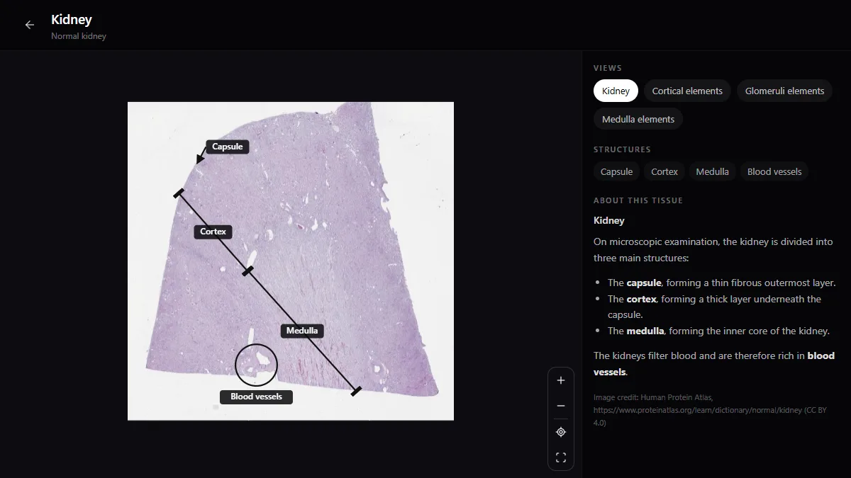
The best online histology atlas lets you zoom a whole slide the way a microscope does and tells you what you are looking at. Free options are now good enough for a whole course: the RadioAnatomy histology atlas for labelled whole-slide sections, Histology Guide and Michigan Histology for virtual microscopy, and the Human Protein Atlas for a vast image collection.
What to look for in a histology atlas
- Whole-slide zoom, from the organ’s architecture down to single cells.
- Labels you can hide, so you can test yourself.
- The standard stain (H&E) plus the special stains your course uses.
- Organisation by tissue and organ, matching your syllabus.
- A viewer that works on a phone, because most revision happens there.
The best free histology atlases online
1. RadioAnatomy
Best for: labelled whole-slide sections, free
The histology atlas shows whole-slide tissue sections with labelled structures, from low power down to single cells. It sits beside the anatomy and radiology atlases, so you can study the same organ in 3D, on CT and under the microscope.
2. Histology Guide
Best for: a complete virtual microscopy course
A free virtual slide box organised like a laboratory manual, with zoomable slides for each chapter of a standard course.
3. Michigan Histology
Best for: a university slide collection with learning objectives
The University of Michigan’s virtual slide collection, free and organised by course unit.
4. Human Protein Atlas
Best for: depth: a very large collection of tissue images
An open research database with images of normal and cancer tissues and a histology dictionary. It is not written as a course, but it is an excellent source of real examples.
5. Kenhub
Best for: histology lessons with quizzes
Articles, labelled images and quizzes that tie histology to anatomy. Part free, part paid.
Pair any atlas with a textbook such as Ross and Pawlina, Junqueira or Wheater for the explanations: the atlas shows you the tissue, the book tells you why it looks that way.
Comparison at a glance
| Atlas | Best for | Cost |
|---|---|---|
| RadioAnatomy | Labelled whole-slide sections | Free |
| Histology Guide | Virtual microscopy course | Free |
| Michigan Histology | University slide collection | Free |
| Human Protein Atlas | Large image database | Free |
| Kenhub | Lessons and quizzes | Part free, part paid |
How to read a histology slide
- Start at low power. Identify the organ’s overall architecture before you zoom in.
- Name the basic tissues you can see: epithelium, connective tissue, muscle, nerve.
- Find the detail that identifies the organ, such as villi, glomeruli or the layers of the wall.
- Know the stain. In H&E, nuclei are blue-purple, and cytoplasm and collagen are pink.
- Compare look-alikes side by side: duodenum against jejunum, parotid against pancreas.
Frequently asked questions
What is the best free histology atlas online?
For labelled whole-slide sections, the RadioAnatomy histology atlas; for a full virtual microscopy course, Histology Guide. Both are free, and they work well together.
How do I study histology effectively?
Look at slides more than you read about them. Go from low to high power, say aloud what identifies the tissue, and test yourself with the labels hidden. Short daily sessions work better than one long one.
Can AI identify histology slides?
AI tools can describe an image, but they confuse look-alike tissues and you cannot check their reasoning. Use a labelled atlas as the reference, and AI only to explain what you have already identified.
RadioAnatomy is our own product, so weigh what we say about it accordingly. We have no affiliate or commercial relationship with the other tools listed.
Get the RadioAnatomy app
Anatomy, radiology and histology in one app, on the App Store and Google Play.