How to Identify Histology Slides, Organ by Organ
To identify a histology slide, work from large to small: the shape of the section at low power, then which of the four basic tissues you see, then the one feature that identifies the organ. Most organs have such a feature, and exams are built around the pairs that look alike.
The method, in four steps
- 1. Lowest power first. Is the organ hollow or solid? Does it have layers, lobules, a cortex and a medulla?
- 2. Name the basic tissues: epithelium, connective tissue, muscle, nervous tissue.
- 3. If there is a lumen, look at its lining: the type of epithelium and what lies under it narrow the answer quickly.
- 4. Find the feature that identifies the organ, and only then zoom in to confirm it.
The feature that identifies each organ
| Organ | What gives it away |
|---|---|
| Oesophagus | Non-keratinised stratified squamous epithelium over a thick muscular wall |
| Stomach | Gastric pits and glands, simple columnar epithelium, no goblet cells |
| Duodenum | Villi, with Brunner’s glands in the submucosa |
| Ileum | Villi, with Peyer’s patches of lymphoid tissue |
| Colon | Straight crypts, no villi, many goblet cells |
| Appendix | A small lumen ringed by lymphoid follicles |
| Liver | Plates of hepatocytes around a central vein, portal triads at the corners |
| Pancreas | Serous acini with pale islets of Langerhans among them |
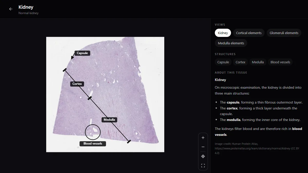
| Kidney | Glomeruli in the cortex; a medulla made only of tubules |
| Thyroid | Follicles filled with pink colloid |
| Lung | Thin-walled alveoli; bronchioles without cartilage |
| Trachea | Ciliated pseudostratified epithelium with goblet cells over hyaline cartilage |
| Skin | Keratinised stratified squamous epithelium, hair follicles and glands in the dermis |
Each link opens the labelled whole-slide section in the free histology atlas, so you can check the feature on a real slide.
The three kinds of muscle
| Muscle | Striations | Nuclei | Other clue |
|---|---|---|---|
| Skeletal | Yes | Many, at the edge of the fibre | Long, unbranched fibres |
| Cardiac | Yes | One or two, central | Branching fibres, intercalated discs |
| Smooth | No | One, central, elongated | Spindle-shaped cells |
Look-alikes that exams use
- Duodenum, jejunum, ileum: Brunner’s glands mean duodenum; Peyer’s patches mean ileum; neither, with tall villi, means jejunum.
- Parotid and pancreas: both are serous, but only the pancreas has islets, and only the parotid has prominent striated ducts.
- Lymph node, spleen, thymus: a lymph node has a capsule, a cortex with follicles and sinuses; the spleen has white pulp around a central arteriole and no cortex or medulla; the thymus has lobules with Hassall’s corpuscles.
- Artery and vein: the artery has a thick muscular wall and a round lumen; the vein has a thin wall and a collapsed lumen.
- Oesophagus and vagina: both have non-keratinised stratified squamous epithelium; the vagina has no glands and no muscularis mucosae.
Know your stains
- Haematoxylin and eosin (H&E): nuclei blue-purple, cytoplasm and collagen pink.
- PAS: glycogen, mucus and basement membranes magenta.
- Masson’s trichrome: collagen blue or green, muscle red.
- Silver stains: reticular fibres black.
How to practise
Cover the label, describe the slide aloud in the four steps, name the organ, then check. Ten minutes a day with a virtual slide box is enough to make the patterns automatic. For more places to practise, see the best free histology atlases online.
Frequently asked questions
How do you identify a tissue under the microscope?
Start at the lowest power to see the overall architecture, decide which of the four basic tissues are present, look at the epithelium lining any lumen, and then find the feature that is specific to the organ, such as glomeruli in the kidney or follicles with colloid in the thyroid.
How do you tell the duodenum, jejunum and ileum apart?
All three have villi. The duodenum has Brunner’s glands in the submucosa, the ileum has Peyer’s patches, and the jejunum has neither.
How do you tell cardiac from skeletal muscle?
Both are striated. Skeletal muscle fibres are long and unbranched with many nuclei at the edge. Cardiac muscle fibres branch, have one or two central nuclei and are joined by intercalated discs.
Get the DecodeMed app
Anatomy, radiology and histology in one app, on the App Store and Google Play.