Knee MRI Anatomy: How to Read a Knee MRI, Plane by Plane
By the DecodeMed Team8 min read
Get the RadioAnato AI app, free:App StoreGoogle PlayAbout RadioAnato AI
Read a knee MRI plane by plane: the sagittal images show the cruciate ligaments, the menisci and the extensor tendons; the coronal images show the collateral ligaments and the meniscal bodies; the axial images show the patellofemoral joint and cartilage. On the usual proton-density (PD) sequences, ligaments, tendons and menisci are dark (low signal), so anything bright inside them is suspicious.
Sequences in one minute
- PD with fat suppression is the workhorse: fluid and bone marrow oedema are bright, fat is dark, ligaments and menisci stay dark.
- T1 shows anatomy and bone marrow: fat and marrow are bright, fluid is dark. Good for fractures and marrow lesions.
- T2 is similar to PD fat-sat for fluid, and helps characterise cysts and effusions.
What each plane is best for
| Plane | Best for | Normal look |
|---|---|---|
| Sagittal | ACL, PCL, anterior and posterior horns of the menisci, quadriceps and patellar tendons | ACL: taut, striated, parallel to the intercondylar roof; PCL: thick, dark, gently curved |
| Coronal | Medial and lateral collateral ligaments, meniscal bodies, cartilage of the condyles | MCL: thin dark band on the medial side; LCL joins the biceps femoris tendon on the fibular head |
| Axial | Patellofemoral joint, patellar cartilage, retinacula, Baker cyst | Smooth cartilage; patella centred in the trochlear groove |
The menisci
The menisci are dark triangles on sagittal and coronal images. On sagittal slices of about 4 mm, the meniscal body looks like a bow tie on two consecutive slices, then the anterior and posterior horns appear as two separate triangles. The posterior horn of the medial meniscus is larger than its anterior horn; the lateral meniscus has horns of similar size. A tear is suggested when abnormal signal reaches an articular surface on at least two images (the “two-slice touch” rule).
The cruciates and collaterals
- ACL: from the inner side of the lateral femoral condyle to the front of the tibial plateau. It should be continuous, taut and roughly parallel to the roof of the intercondylar notch (Blumensaat line). A wavy, discontinuous or horizontal ACL suggests a tear.
- PCL: thicker and uniformly dark, curving from the medial condyle to the back of the tibia; on sagittal images it looks like a hockey stick.
- MCL: a thin dark band from the medial epicondyle to the tibia, blending with the joint capsule and the medial meniscus.
- LCL: from the lateral epicondyle to the fibular head, where it joins the biceps femoris tendon; the popliteus tendon runs nearby.
A checklist to read any knee MRI
- 1. Effusion and Baker cyst (between the medial head of gastrocnemius and semimembranosus).
- 2. Bone marrow: oedema, contusions, fractures.
- 3. Menisci: medial, then lateral, horn by horn.
- 4. Cruciate ligaments, then 5. collaterals.
- 6. Extensor mechanism: quadriceps tendon, patella, patellar tendon.
- 7. Cartilage of the patella, trochlea and both condyles.
- 8. Muscles, vessels and nerves in the popliteal fossa.
Learn it on a labelled knee MRI
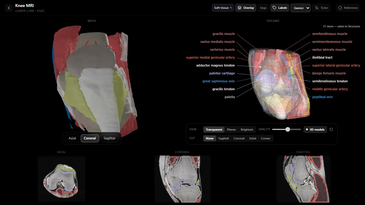
The radiology atlas has a knee MRI in which every structure is rebuilt as a 3D model and labelled on the volume: the semitendinosus, semimembranosus, gracilis and sartorius, the patellar cartilage, the genicular arteries, the popliteal vein and more. Below, the axial, coronal and sagittal slices show the same structures outlined, so you can move from the 3D model to the plane you will read in clinic.
Study it in RadioAnato AI
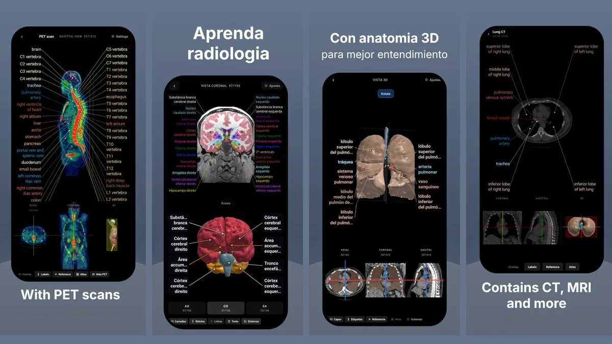
RadioAnato AI, the DecodeMed app, puts 3D anatomy, CT and MRI studies, histology slides and an AI tutor that answers on them in your pocket. Free to download for iPhone, iPad and Android.
For the basics of signal, read T1 vs T2 MRI; for other studies, the best radiology anatomy atlases.
Frequently asked questions
Which MRI plane is best to see the ACL?
The sagittal plane, where the ACL appears as a taut, striated dark band running parallel to the roof of the intercondylar notch. Coronal and axial images help confirm a tear.
What does a normal meniscus look like on MRI?
A uniformly dark triangle. On sagittal slices the body forms a bow tie on two consecutive images; signal that reaches an articular surface on two images suggests a tear.
Why are ligaments dark on MRI?
They are made of tightly packed collagen with very little free water, so they give almost no signal on PD, T1 and T2 sequences.
What is the best sequence for knee MRI?
Proton density with fat suppression in all three planes, plus T1 for anatomy and bone marrow. Protocols vary between centres.
Get RadioAnato AI, the DecodeMed app
Anatomy, radiology and histology in one app, on the App Store and Google Play.
About RadioAnato AI