RBBB vs LBBB on the ECG: How to Tell Them Apart
By the DecodeMed Team7 min read
Get the RadioAnato AI app, free:App StoreGoogle PlayAbout RadioAnato AI
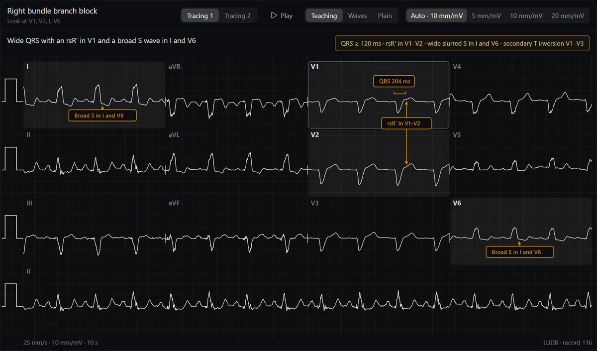
Both blocks widen the QRS to 120 ms or more; look at V1 to tell them apart. In right bundle branch block (RBBB) V1 shows an rsR′ (“M” shape) and I and V6 have a broad, slurred S wave. In left bundle branch block (LBBB) V1 is mostly negative (QS or rS) and I, aVL and V6 show a broad, notched or monophasic R with no septal q wave.
Why the QRS widens
Normally both ventricles are activated at the same time through the right and left bundle branches. When one branch is blocked, its ventricle is activated late, slowly, muscle cell to muscle cell, from the other side. That late activation adds a broad extra deflection to the QRS, pointing toward the late ventricle: rightward and anterior in RBBB (the R′ in V1), leftward and posterior in LBBB (the tall R in V6).
Side-by-side criteria
| RBBB | LBBB | |
|---|---|---|
| QRS duration | ≥ 120 ms (110–119 ms = incomplete) | ≥ 120 ms |
| V1–V2 | rsR′, “M” or “rabbit ears” | QS or rS, broad and negative |
| I, aVL, V5–V6 | Broad, slurred S wave | Broad, notched or monophasic R; no septal q |
| ST–T | T inverted in V1–V3 (secondary) | Discordant: opposite the main QRS deflection |
| Axis | Usually normal | Often normal or left |
| Clinical meaning | Can be normal; also PE, ASD, right heart strain | Almost always underlying heart disease |
The WiLLiaM MaRRoW trick
Look at V1 and V6. In LBBB, V1 looks like a W and V6 like an M: WiLLiaM. In RBBB, V1 looks like an M and V6 like a W: MaRRoW. It is a memory aid, not a criterion; real tracings are often not textbook-shaped, so confirm with the QRS width and the leads above.
Causes
- RBBB: can be found in healthy people; also pulmonary embolism, atrial septal defect, right ventricular strain, ischaemic heart disease and after cardiac surgery.
- LBBB: hypertension, coronary artery disease, dilated cardiomyopathy, aortic stenosis, degeneration of the conduction system. A new LBBB deserves an evaluation of the heart.
LBBB and myocardial infarction
Because LBBB already shifts the ST segment, the usual STEMI criteria do not apply. Current guidelines no longer treat a new LBBB alone as a STEMI equivalent; instead, use the Sgarbossa criteria (concordant ST elevation ≥ 1 mm, concordant ST depression ≥ 1 mm in V1–V3) or the modified version based on the ST/S ratio, together with the clinical picture. RBBB does not hide ST elevation, so a STEMI can be read through it.
Practise on real tracings
The ECG atlas shows bundle branch blocks on real 12-lead recordings. The Teaching view puts arrows on the rsR′ and the broad S wave and lists the criteria; switch to Plain and find them yourself.
Study it in RadioAnato AI
RadioAnato AI, the DecodeMed app, puts 3D anatomy, CT and MRI studies, histology slides and an AI tutor that answers on them in your pocket. Free to download for iPhone, iPad and Android.
Read every ECG in the same order: see how to learn ECG from scratch, normal ECG values and how to calculate the axis.
Frequently asked questions
How do you tell RBBB from LBBB?
Look at V1. An rsR′ (M shape) with a positive terminal deflection is RBBB; a broad, mostly negative QS or rS is LBBB. Both need a QRS of 120 ms or more.
Is right bundle branch block dangerous?
An isolated RBBB is often found in healthy people. It matters when it is new or comes with symptoms, for example in pulmonary embolism.
Can you diagnose a myocardial infarction with LBBB?
Not with the usual ST criteria. Use the Sgarbossa or modified Sgarbossa criteria together with symptoms, troponin and, if needed, urgent angiography.
What is the WiLLiaM MaRRoW mnemonic?
In LBBB, V1 looks like a W and V6 like an M; in RBBB, V1 looks like an M and V6 like a W. Use it as a reminder, then confirm with the criteria.
Get RadioAnato AI, the DecodeMed app
Anatomy, radiology and histology in one app, on the App Store and Google Play.
About RadioAnato AI