How to Learn ECG with AI: Vectors, Real Tracings and Socratic Drills
By the DecodeMed Team7 min read
Get the RadioAnato AI app, free:App StoreGoogle PlayAbout RadioAnato AI
The right way to learn ECG with AI is never to upload a photo of a paper strip and ask a chatbot to read it for you. Instead, combine the DecodeMed ECG Atlas (real cardiologist-annotated 12-lead tracings with Plain, Waves and Teaching modes) with RadioAnato AI (the DecodeMed app whose AI tutor maps each lead onto the 3D heart, coronary arteries, CT scan and cardiac histology). Read the tracing yourself first, check the wave markers, and use the AI tutor to deduce waveforms from 3D electrical vectors.
Why general chatbots misread ECG photos—and why RadioAnato AI + DecodeMed ECG Atlas works
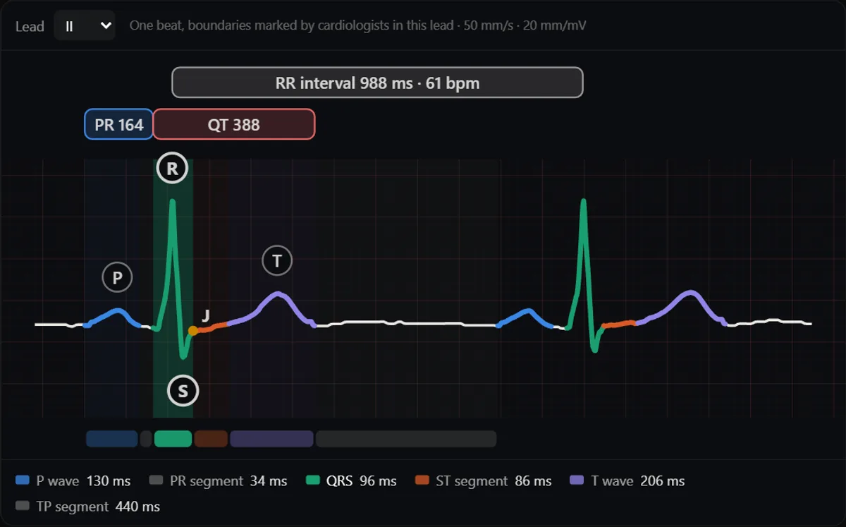
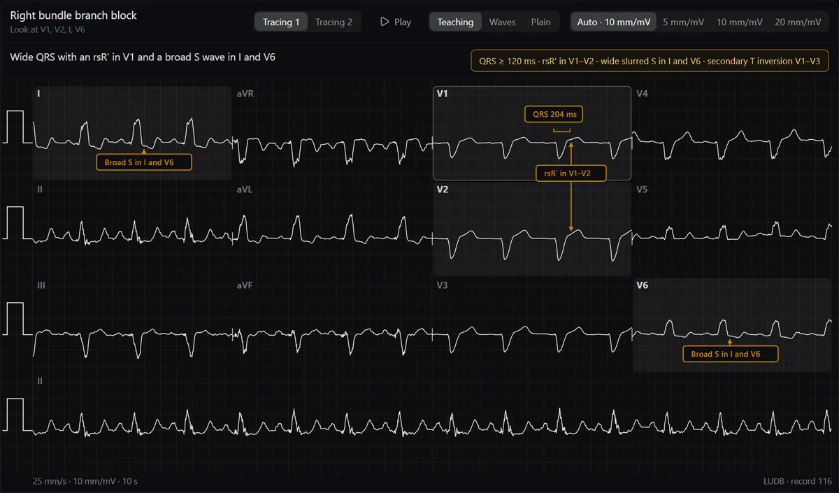
General vision chatbots cannot measure a 40 ms small square reliably on a skewed phone photo, and they frequently hallucinate ST elevation or miss subtle P waves. Worse, letting a chatbot interpret the tracing robs you of the visual pattern recognition you need on the wards. By pairing the DecodeMed ECG Atlas with RadioAnato AI, you practise on real 12-lead recordings where cardiologists marked every P, QRS, ST and T wave, while the AI tutor shows you the 3D heart wall and coronary artery that each lead looks at.
| Learning task | Ineffective chatbot habit | High-yield workflow with RadioAnato AI & DecodeMed |
|---|---|---|
| Reading a 12-lead ECG | Uploading a phone photo of an ECG to a general chatbot | Reading a real tracing in Plain mode in the DecodeMed ECG Atlas, then checking Waves and Teaching mode |
| Understanding leads | Memorising lead groups as arbitrary lists | Asking the RadioAnato AI tutor to show the 3D heart and coronary arteries so you see which wall each lead faces |
| Learning criteria | Reading passive text summaries of bundle branch blocks | Having the AI tutor deduce the V1 and V6 waveform with you from the 3D cardiac depolarization vector |
| Differential diagnosis | Studying arrhythmias one by one in isolation | Running AI comparison drills (Mobitz I vs II, pericarditis vs STEMI, VT vs SVT with aberrancy) |
The 4-step workflow to learn ECG with RadioAnato AI
- Step 1 — Map the 12 leads onto the 3D heart in RadioAnato AI (5 min). Open the heart in RadioAnato AI on iPhone, iPad or Android (or in the free web 3D anatomy atlas). Ask the AI tutor to highlight the right ventricle anteriorly (V1–V2), the interventricular septum (V3–V4), the lateral left ventricular wall (V5–V6, I, aVL) and the inferior wall resting on the diaphragm (II, III, aVF), plus the coronary arteries supplying each wall.
- Step 2 — Write a blind 6-line reading in the DecodeMed ECG Atlas (5 min). Open a case in the free DecodeMed ECG Atlas and switch to Plain view. Write down: (1) Rate, (2) Rhythm, (3) Axis, (4) PR, QRS and QT intervals, (5) Wave morphology and ST segment, and (6) Diagnosis. Switch to Waves and Teaching view to grade your measurements.
- Step 3 — Ask AI to challenge your vector reasoning (10 min). Use the prompts below in RadioAnato AI (or alongside your AI assistant). Instead of asking “what is RBBB?”, make the AI walk you from the blocked bundle to the 3D direction of the electrical vector and ask *you* whether V1 will record a positive or negative deflection.
- Step 4 — Drill look-alike differentials (5 min). Quiz yourself on the two ECG patterns you most easily confuse and turn the tie-breaker criterion into an Anki card.
Study it in RadioAnato AI
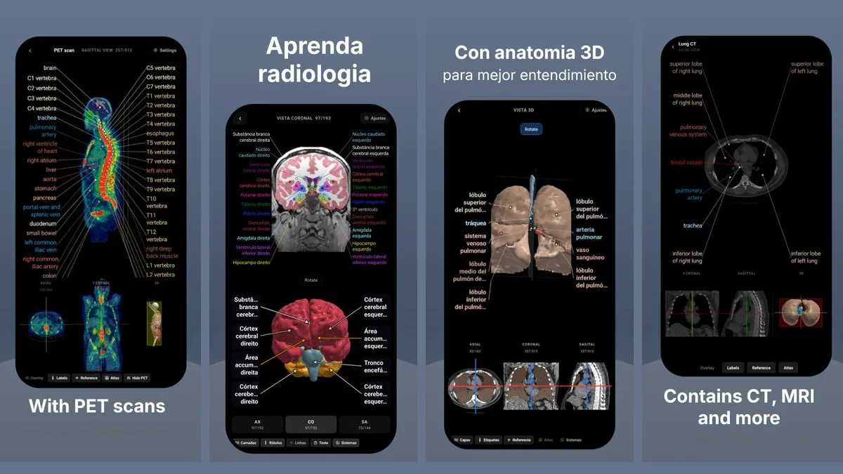
RadioAnato AI, the DecodeMed app, puts 3D anatomy, CT and MRI studies, histology slides and an AI tutor that answers on them in your pocket. Free to download for iPhone, iPad and Android.
Prompts to use in RadioAnato AI and your ECG study
3D coronary territory & lead check (ask in RadioAnato AI)
Show me the 3D heart and the [left anterior descending / right coronary / left circumflex] artery. Explain which ECG leads face the myocardium supplied by this artery and what reciprocal changes appear on the opposite wall.
Vector-to-wave Socratic deduction (stop memorising shapes)
Act as a cardiac electrophysiology tutor. Help me deduce the 12-lead ECG appearance of [left bundle branch block / inferior STEMI / right ventricular hypertrophy] from first principles. Do not give me the criteria list at once. Ask me step by step: (1) which way the depolarization or injury vector points in 3D, (2) what lead V1 sees and why, and (3) what lead V6 or lead II sees and why.
Critique my 6-step ECG interpretation
I just read a practice ECG on my own. Here are my findings: [Rate: 42 bpm, regular; P waves: 85 bpm, marching through regularly; PR interval: variable, no relationship between P and QRS; QRS: 140 ms]. Ask me: (1) what my primary diagnosis is, (2) where the escape pacemaker is likely located based on the QRS width, and (3) what clinical causes I should consider.
Look-alike tie-breaker drill
Quiz me on the ECG differences between [acute pericarditis, early repolarisation and acute ST-elevation myocardial infarction]. Give me five short written ECG descriptions, one at a time, and ask me to name the most likely pattern and the single strongest ECG feature that separates it from the other two.
Build your foundation with DecodeMed’s free ECG resources
- Practise on real annotated 12-lead tracings in the free DecodeMed ECG Atlas and download RadioAnato AI on iOS and Android.
- Follow a fixed reading order every time: see how to learn to read an ECG from scratch and normal ECG values, waves and intervals.
- Master the two quick calculations: how to calculate heart rate on an ECG, how to calculate the electrical axis and ECG practice cases with answers.
To compare the tools themselves, read the best AI tools to learn ECG interpretation and the best apps to learn ECG.
Frequently asked questions
What is the best way to learn ECG with AI?
Combine the DecodeMed ECG Atlas (where real 12-lead tracings have every wave coloured and explained by cardiologists in Teaching, Waves and Plain views) with RadioAnato AI (whose AI tutor shows the 3D heart, coronary arteries and cardiac histology behind each lead).
Why shouldn’t I upload an ECG photo to ChatGPT to learn?
General vision models cannot reliably calibrate paper speed and voltage grid squares on a photo, often missing subtle P waves or inventing ST shifts. Always read real calibrated tracings in the DecodeMed ECG Atlas instead.
How can RadioAnato AI help me stop memorising ECG patterns by rote?
Ask the RadioAnato AI tutor to show the 3D heart, its chambers, coronary arteries and conduction system, then trace the direction of the electrical vector in 3D so you can predict whether each lead records an upward or downward wave.
Get RadioAnato AI, the DecodeMed app
Anatomy, radiology and histology in one app, on the App Store and Google Play.
About RadioAnato AI