Brain MRI Anatomy: Basal Ganglia, Ventricles and Internal Capsule Step by Step
By the DecodeMed Team7 min read
Get the RadioAnato AI app, free:App StoreGoogle PlayAbout RadioAnato AI
The fastest way to orient yourself on an axial brain MRI is to find the CSF spaces in the midline first—the frontal horns of the lateral ventricles and the slit-like third ventricle—and then read the deep grey nuclei arranged around the V-shaped white matter of the internal capsule. You can scroll this exact volume with 3D reconstructions in the free DecodeMed radiology atlas or ask the AI tutor in RadioAnato AI to highlight the caudate, putamen, globus pallidus and thalamus in 3D.
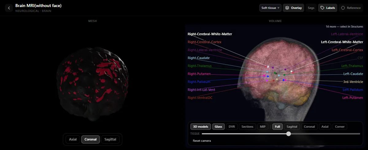
Step 1: Confirm the sequence and the midline
- Check the CSF in the ventricles first. If the ventricular fluid is dark and white matter is brighter than cortical grey matter, you are looking at a T1-weighted image (the classic “anatomical” sequence). If the CSF is bright white, you are on T2. See T1 vs T2 MRI: how to tell them apart.
- Trace the midline from anterior to posterior: the falx cerebri between the frontal lobes, the septum pellucidum separating the two frontal horns, and the narrow third ventricle between the two thalami.
Step 2: The “star” level — basal ganglia and internal capsule
Scroll to the axial slice where both the frontal horns of the lateral ventricles and the third ventricle are visible together. Everything important in the deep brain sits around a boomerang-shaped band of white matter called the internal capsule:
| Structure | Where it sits on the axial slice | Key clinical role |
|---|---|---|
| Caudate nucleus (head) | Bulges into the lateral wall of the frontal horn, anterior and medial to the internal capsule | Atrophies in Huntington disease, enlarging the frontal horns |
| Internal capsule (anterior limb, genu, posterior limb) | V-shaped white-matter highway pointing medially toward the third ventricle | The posterior limb carries the corticospinal tract; a tiny lacunar infarct here causes pure motor hemiparesis |
| Lentiform nucleus (putamen + globus pallidus) | Wedge-shaped grey mass tucked lateral to the internal capsule (putamen is outer, pallidus is inner) | Common site of hypertensive intracerebral haemorrhage |
| Thalamus | Large oval grey nucleus medial to the posterior limb of the internal capsule, flanking the third ventricle | Main sensory relay to the cortex |
| Insular cortex & Sylvian fissure | Lateral to the lentiform nucleus, separated by the external capsule and claustrum | Supplied by the middle cerebral artery (MCA); loss of the insular ribbon is an early sign of stroke |
Step 3: Follow the ventricular system from top to bottom
- Bodies and frontal horns of the lateral ventricles sit high beneath the corpus callosum, separated by the septum pellucidum.
- Trigone (atrium), occipital and temporal horns sweep posteriorly and inferiorly into the occipital and temporal lobes.
- Foramina of Monro connect the lateral ventricles to the midline third ventricle between the thalami.
- Cerebral aqueduct (of Sylvius) runs through the midbrain into the fourth ventricle, which sits between the pons/medulla anteriorly and the cerebellum posteriorly.
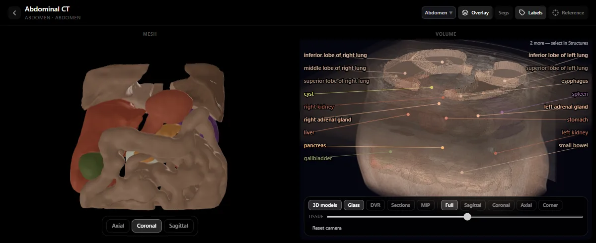
Study it in RadioAnato AI
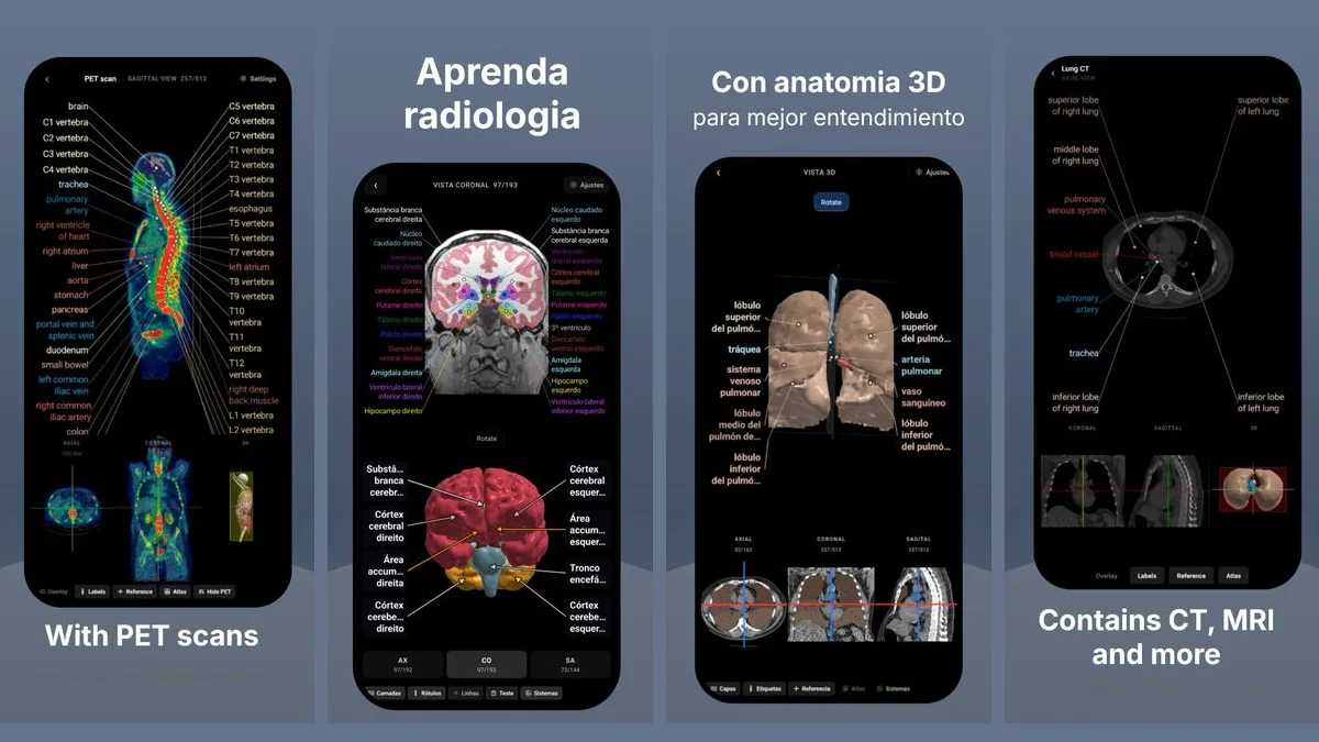
RadioAnato AI, the DecodeMed app, puts 3D anatomy, CT and MRI studies, histology slides and an AI tutor that answers on them in your pocket. Free to download for iPhone, iPad and Android.
Step 4: Three levels above and below the basal ganglia
- High convexity (centrum semiovale): above the ventricles, where the central sulcus separates the precentral gyrus (motor strip) from the postcentral gyrus. Look for the “omega sign” of the hand motor area.
- Midbrain level: shaped like Mickey Mouse’s ears (the cerebral peduncles anteriorly) with the cerebral aqueduct and quadrigeminal cistern posteriorly.
- Pons and cerebellopontine angle: the belly of the pons anteriorly, the fourth ventricle behind it, and the middle cerebellar peduncles reaching into the cerebellum.
Open the labelled brain MRI volume in the free radiology atlas to rotate the 3D deep grey nuclei while scrolling the slices. For more cross-sectional guides, read chest CT anatomy, how to read an abdominal CT and how to learn radiology with AI.
Frequently asked questions
How do you tell the caudate, putamen and thalamus apart on an axial brain MRI?
Find the V-shaped white-matter band of the internal capsule first. The caudate head is anterior and medial (hugging the frontal horn of the lateral ventricle), the thalamus is posterior and medial (hugging the third ventricle), and the wedge-shaped lentiform nucleus (putamen and globus pallidus) sits laterally in the crook of the V.
Which MRI sequence is best for learning normal brain anatomy?
T1-weighted MRI is best for normal anatomy because CSF is dark, grey matter is grey and white matter is lighter grey, matching how the brain looks in neuroanatomy lab.
Where can I practise scrolling a labelled brain MRI for free?
Open the DecodeMed radiology atlas at decodemed.app/rad-atlas in your browser or download RadioAnato AI for free on iPhone, iPad and Android to scroll a real brain MRI with the cortex, white matter, ventricles, caudate, putamen, globus pallidus and thalamus labelled in 2D and 3D.
Get RadioAnato AI, the DecodeMed app
Anatomy, radiology and histology in one app, on the App Store and Google Play.
About RadioAnato AI